Fever, Chills, and Nonhealing Perineal Ulcer in a 51-Year-Old Woman
A 51-year-old woman has had a worsening perineal ulcer for months despite treatment. She now has a new productive cough and mild dysuria. What’s the diagnosis? Case by Gustavo Freitas et al.
Editor's Note : The Case Challenge series includes difficult-to-diagnose conditions, some of which are not frequently encountered by most clinicians, but are nonetheless important to accurately recognize. Test your diagnostic and treatment skills using the following patient scenario and corresponding questions.
If you have a case that you would like to suggest for a future Case Challenge, please email us at ccsuggestions@medscape.com with the subject line "Case Challenge Suggestion." We look forward to hearing from you. Background and Initial Presentation A 51-year-old woman with a past medical history of herpes simplex virus type 2 (HSV-2) infection, pneumocystis pneumonia, and HIV infection presents to the emergency department with fever, chills, and fatigue following a wound care visit for a chronic, nonhealing perineal ulcer unresponsive to oral acyclovir .
She reports that the wounds have been present for 3 months and have progressively worsened. Two months prior, she was prescribed valacyclovir for suspected HSV reactivation without improvement and was subsequently switched to acyclovir. She also reports poor oral intake and a new productive cough.
Her current medications include acyclovir and prophylactic rifabutin and sulfamethoxazole-trimethoprim . She is not receiving antiretroviral therapy. In the emergency department, the patient reports pain rated 5 out of 10, along with associated constipation and mild dysuria.
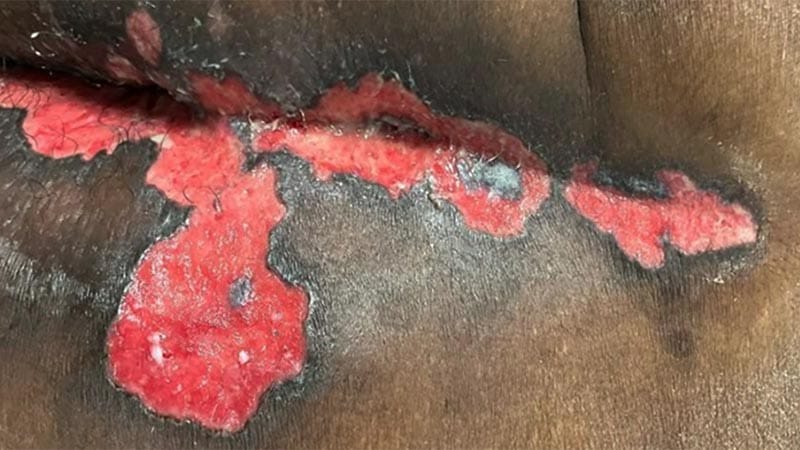
Physical Examination On physical examination, her oral temperature is 100.9 °F (38.2° C), heart rate is 119 beats/min, blood pressure is 140/83 mm Hg, and oxygen saturation is 98% on room air. Dermatologic examination reveals monomorphic pink to red ulcerative plaques on the right buttock, extending into the gluteal cleft and inguinal folds (Figure 1).
Lesions are not draining, bleeding, or malodorous. The patient is admitted meeting systemic inflammatory response syndrome (SIRS) criteria with concern of sepsis in the setting of superimposed cellulitis . Figure 1 .
The patient's gluteal cleft with a pink to red ulcerative plaques with a hyperpigmented border. Initial evaluation prioritizes identification of systemic infection in a patient meeting SIRS criteria. Bacterial and viral culture are crucial to identify infectious source and guide antimicrobial therapy. [1,2] In addition, when acyclovir resistance is suspected, the lesion should be cultured and any viral isolate sent for susceptibility testing.
HSV PCR is the preferred diagnostic test for mucocutaneous lesions but does not provide susceptibility data. [3] Ultrasonography, urinalysis, and CT scan are reasonable adjuvant studies depending on patient presentation. Although these studies can be useful in identifying an underlying etiology, the patient's initial presentation raises concern for impending systemic decompensation from an infection, making identification of the infectious source the immediate priority.
Workup Given fever, tachycardia, and immunocompromised status, a broad infectious workup was initiated. Blood and wound cultures, urinalysis, STI testing, COVID-19 , cytomegalovirus NAAT, HSV , and varicella zoster virus (VZV) polymerase chain reaction (PCR), chest x-ray, and punch biopsy were performed.
Laboratory studies showed a white blood cell count of 2.8 × 10³/μL, procalcitonin level of 0.17 ng/mL, and an absolute CD4 count of 5 cells/μL. Leukopenia and a low CD4 count were consistent with HIV-associated immunosuppression. Procalcitonin is used to differentiate between bacterial and viral infections, with 0.17 ng/mL supporting a lower suspicion for bacterial infection.
Nonetheless, current sepsis guidelines suggest using clinical evaluation alone, rather than procalcitonin plus clinical evaluation, to decide whether to start antimicrobial therapy. [2] Blood cultures showed no growth. Wound cultures were negative for aerobic and anaerobic bacteria and mycobacteria; however, they demonstrated scant numbers of Candida albicans , thought to represent colonization.
Negative wound and blood cultures further supported a nonbacterial etiology. Urinalysis was notable for pyuria and negative for nitrites. Chlamydia and gonorrhea nucleic acid amplification test (NAAT) and syphilis testing were negative, making an STI less likely.
SARS-CoV-2 testing was positive, which was thought to contribute to the patient's cough; however, the condition was self-limited during her prolonged hospital course. Chest x-ray demonstrated no evidence of acute cardiopulmonary processes. HSV-2 NAAT test results were positive, while HSV-1, VZV NAAT were negative.
Cytomegalovirus NAAT was negative, and IgM and IgG were undetectable. Antiviral susceptibility testing was sent to an outside laboratory; results were pending at the time of this evaluation. Punch biopsy demonstrated ulceration and mixed dermal inflammatory infiltrate.
HSV immunostaining was positive. Human herpesvirus-8 (HHV8) and varicella immunostains were negative. Periodic acid–Schiff stain showed no definitive fungal elements (Figure 2).
Figure 2 . Hematoxylin and eosin staining, 2x and 4x, and immunostaining positive for HSV and negative for VZV and HHV8. In an immunocompromised patient with recurrent and prolonged genital ulcers, HSV infection is an important diagnostic consideration.
Acyclovir resistance occurs in 3.5%–7% of individuals with HIV infection. [4,5] Immunocompromise, chronic progressive ulceration, positive HSV test results, and failure to respond to appropriate antiviral therapy are highly suggestive of acyclovir -resistant HSV infection. Current guidelines advise suspecting resistance when lesions do not begin to resolve within 7-10 days of therapy. [3] A plaque reduction assay can confirm antiviral resistance. [3,4] Antiviral susceptibility testing performed by the outside laboratory confirmed acyclovir resistance.
Diagnosis Acyclovir -resistant HSV infection is rare in immunocompetent individuals, occurring in less than 1% of patients. [5,6] However, resistance is not uncommon in immunocompromised individuals, particularly among those on prolonged acyclovir prophylaxis. [3] Other risk factors include suboptimal dosing, profound immunosuppression, and ongoing viral replication.
Perirectal lesions are more likely than lesions at other sites to be acyclovir resistant. [3] High-risk patients include hematopoietic stem cell transplant (HSCT) recipients, patients with multiple myeloma, and patients with HIV . [5,7] Cutaneous CMV infection is rare and may present with anogenital ulcerations, most frequently in patients with CD4 counts below 50 cells/uL.
Anogenital ulcerations may test positive for both CMV and HSV, although often resolving with HSV-directed antivirals alone, indicating that HSV is the principal pathogen. CMV infection is treated with ganciclovir or valganciclovir . [8] In immunocompromised patients with ulcerations not responding to acyclovir, CMV PCR or biopsy may be considered.
CMV PCR and serum antibodies were negative in this patient. Although HIV is a risk factor for cutaneous tuberculosis , the patient has no reported history of risk factors associated with exposure, including occupational exposure, travel to endemic areas, or injection drug use. Negative acid-fast bacilli culture and stain further reduce suspicion for this diagnosis.
Pyoderma gangrenosum (PG) can also present with eroded pink plaques. PG is often associated with inflammatory conditions, including inflammatory bowel disease, which is not reported in this patient. Histopathologically, PG is more commonly associated with a neutrophilic infiltrate on biopsy rather than the mixed inflammatory infiltrate reported in this patient, making this diagnosis less likely. [9] Although the most common morphology of HSV infection is grouped vesicles and “punched-out” lesions on an erythematous base, this morphology is not suggestive of acyclovir-resistant HSV . [3] In cases of antiviral resistance, HSV lesions can present atypically with verrucous, hypertrophic, or ulcerative appearance.
These morphologies reflect chronic, poorly controlled viral replication in immunocompromised hosts and occur with resistant disease, but they are not specific to resistance; hypertrophic genital HSV infection in people with HIV can mimic neoplasia and requires biopsy for diagnosis. [3] In rare cases, granulomatous lesions and necrotizing cutaneous lesions may occur. [10,11,12] Annular lesions with a trailing collarette suggest erythema annulare centrifugum , which commonly presents on the thighs and buttocks.
In contrast, targetoid lesions with a dusky center are characteristic of erythema multiforme , which is immune-mediated and self-limiting. Discussion In patients with nonresistant HSV infection, recurrent HSV episodes typically last 5-10 days if antiviral therapy is not given. [3] Immunocompromised patients are at higher risk for more extensive and prolonged reactivation of the virus. [3,4] Atypical presentations may require further diagnostic testing to rule out cytomegalovirus infection and cutaneous tuberculosis .
A punch biopsy and PCR for HSV and CMV, and wound culture for acid-fast bacilli to rule out tuberculosis, may be performed. HSV culture and susceptibility phenotyping can confirm acyclovir resistance. [3] However, in serious cases, including high risk for dissemination, empiric treatment should be started before results are available. [13] Acyclovir resistance is thought to occur through thymidine kinase-deficient mutants of the HSV-2 virus, altered substrate specificity of thymidine kinase, or alteration in DNA polymerase. [5,7] Acyclovir, valacyclovir , famciclovir , and ganciclovir are acyclic guanosine analogues that are activated by viral thymidine kinase and inhibit DNA polymerase.
When thymidine kinase is absent or DNA polymerase is altered, acyclovir and its structural analogues are no longer efficacious, resulting in antiviral resistance. [5] All acyclovir-resistant strains are resistant to valacyclovir, and most are resistant to famciclovir. [3] Foscarnet is a pyrophosphate analogue that binds to the pyrophosphate-binding site of the viral DNA polymerase, inhibiting further nucleotide incorporation. [5] Foscarnet does not require thymidine kinase for its antiviral effect, allowing activity against resistant HSV strains. [14] Foscarnet is available topically or intravenously because of its poor bioavailability.
For IV administration, hospital admission is typically required. [14] Close monitoring of complete blood count, serum electrolytes, and renal function tests is required given the risk of foscarnet-induced anemia and granulocytopenia, electrolyte disturbances, and nephrotoxicity. [15] Dual resistance to acyclovir and foscarnet arises from mutations in the viral DNA polymerase; thymidine kinase mutations alone do not confer foscarnet resistance. [5,14] Cidofovir , an acyclic nucleoside phosphonate, is another commonly used antiviral to treat acyclovir-resistant HSV.
Cidofovir's active metabolite inhibits DNA polymerase without requiring viral thymidine kinase. [5] Cidofovir is not considered first-line for acyclovir-resistant HSV cases, as it has a risk for nephrotoxicity and requires extensive monitoring, with one retrospective study demonstrating nephrotoxicity in 25% of hematopoietic cell transplant recipients. [16] Pritelivir is an oral HSV helicase-primase inhibitor. [3,14] In October 2026, the FDA approved pritelivir for immunocompromised adults with mucocutaneous HSV lesions that have not responded to acyclovir, valacyclovir, or famciclovir. [17] The HHS OI Guidelines, last updated before this approval, continue to list IV foscarnet as the preferred therapy for acyclovir-resistant HSV. [3] After a plaque reduction assay confirmed acyclovir-resistant HSV, the patient in this case was readmitted to the hospital for a 14-day course of foscarnet.
Throughout her hospital course, the perineal ulcers and pain improved. Acyclovir -resistant HSV is associated with increased morbidity in immunocompromised individuals. A retrospective study of HSCT recipients found a 100-day mortality rate of 64% in those with active acyclovir-resistant HSV infection compared with 39% in those with active acyclovir-sensitive HSV infection. [18] A delay in adequate treatment may result in dissemination to visceral organs, resulting in meningitis , encephalitis , hepatitis , and pneumonitis. [3] Postherpetic neuralgia is associated with herpes zoster reactivation.
Kaposi sarcoma -associated lesions are a complication of HHV-8 infection. Autoimmune blistering diseases, including bullous pemphigoid and pemphigus vulgaris , are not associated with acyclovir-resistant HSV. Upon administration of foscarnet , lesions should improve within 5-7 days.
If no improvement is observed, consider potential resistance. In a retrospective cohort, 15 of 31 treated episodes healed; among episodes that resolved, the median time to healing was 38 days. [14] At discharge, topical cidofovir was initiated in this patient. Her ulcer continues to improve, although the wounds are not yet fully healed.
The patient continues to decline antiretroviral therapy for her HIV infection. Referrals to the wound clinic, infectious disease clinic, and dermatology have been made. Editor's Note: This article was created using several editorial tools, including generative AI models, as part of the process.
Human review and editing of this content were performed prior to publication.
Article text via FreeNewsAPI. Rights remain with Medscape.
Read on publisher site → Opens Medscape in a new tab