Tokyo develops 3D microscopy at 19.2 frames per second — News-Medical
Researchers at the University of Tokyo have developed a high-speed microscopy method that makes it possible to record dynamic processes inside living cells in three dimensions. The new system produces a...
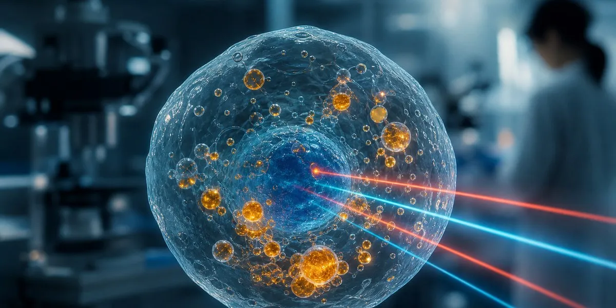
Researchers at the University of Tokyo have developed a high-speed microscopy method that makes it possible to record dynamic processes inside living cells in three dimensions. The new system produces a complete 3D image every 52 milliseconds, or 19.2 images per second, News-Medical reports .
Faster than previous methods The technology is called video-rate mid-infrared photothermal optical diffraction tomography, or MIP-ODT. It uses mid-infrared light, which causes chemical bonds in molecules to vibrate. Different molecules have their own frequency signatures, allowing researchers to create a map of a cell's chemical composition without using dyes.
According to the study's authors, previous methods of three-dimensional mid-infrared photothermal tomography operated at less than 0.05 images per second. The new approach is approximately 400 times faster. To achieve this, the team used a spatial light modulator that enables rapid changes to the angle of sample illumination and optimized the signal-to-noise ratio during each capture.
More current news is available on the UA.News Telegram channel Telegram . Observing the movement of structures inside a cell Using MIP-ODT, scientists observed lipid droplets in a cell and tracked their movement in three-dimensional space. Lipid droplets are involved in energy regulation and lipid storage.
The authors note that 3D visualization made it possible to see features of their transport that are more difficult to detect in two-dimensional imaging. The study's first author, Masato Fukushima, said the method transforms a technology that was mainly used to observe relatively static samples into a tool for quantitatively studying rapid biological processes in three dimensions.
The team is also considering using the method to study biomolecular condensates — membraneless cellular structures made of proteins and nucleic acids.
Article text via FreeNewsAPI. Rights remain with UA.NEWS.
Read on publisher site → Opens UA.NEWS in a new tab